We greatly appreciate the questions that our readers submit. We recently received a collection of questions about lung biopsy.
Why is Lung Biopsy Important in Pulmonary Fibrosis?
Pulmonary fibrosis is a general term that describes scarring in the lungs. There are many lung diseases that have lung scarring as a feature. Determining the specific lung disease is critically important in order to provide accurate information about prognosis and to guide treatment plans.
 For many patients, the history, physical exam, laboratory testing, pulmonary function testing and most importantly the high resolution lung CT scan provides enough information to arrive at a confident diagnosis. However, for other patients the diagnosis remains unclear. In these patients a lung biopsy should be considered.
Some readers have asked why not just try a course of treatment with immunosuppressive medications (such as prednisone). In general, experts for several reasons do not advise this. The most important reason not to embark on a trial of prednisone is that prednisone has many risks such as increased risk of infection, osteoporosis and bone fractures. Furthermore, in a recent landmark study of immunosuppression for IPF, patients receiving immunosuppression had worse outcomes.
What are the Risks of Lung Biopsy?
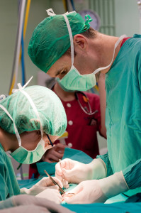
The lung biopsy procedure is called VATS (Video Assisted Thoracoscopic Surgery). This type of procedure is done under general anesthesia. 2 small holes are made in the chest wall and tools are inserted into the chest. 2 or 3 small strips of lung tissue from 2 different lobes of the lung are then taken. A chest tube is then inserted into the chest to re-expand the lung.
The risks of this procedure are those associated with anesthesia (problems inserting a breathing tube, low blood pressure, stroke and even death). Fortunately anesthesia risks are very small with modern techniques. Risks of the surgery include bleeding, infection, and air leak from the lung biopsy site. Most patients spend one or two nights in the hospital. If there is a prolonged air leak then a long hospital stay is required. All patients experience some degree of postoperative pain at the chest tube site (the tube used to re-expand the lung). For the majority of patients this takes a few weeks to resolve, though for some patients pain may linger for months.
Once the lung biopsy is completed the lung tissue needs to be reviewed by a pathologist with special training in lung diseases. The features that distinguish IPF from the other types of pulmonary fibrosis can be subtle to the non-specialist’s eye. If your lung biopsy was done in a smaller hospital you should request in advance to have a second opinion from an expert pathologist (these super-specialized lung pathologists usually are at major universities). It is worth coordinating this in advance. If your pulmonologist or surgeon can’t make this happen then consider finding a different team.
Does Every Patient Need a Surgical Lung Biopsy?
Absolutely not. Many patients will have high resolution CT scan results combined with the rest of the information that allows your doctor to make a confident diagnosis of IPF. Certain features are considered atypical and point to other non-IPF fibrotic diagnoses. These features include upper lung predominance (more involvement of the top of the lungs than the bottom), ground glass and air trapping (ground glass is a hazy quality to the lung images and air trapping signifies air flow obstruction). Lack of honeycomb change also makes IPF less likely. Age less than 50 makes IPF much less likely and suggests alternative diagnoses are more likely. Under these conditions a biopsy may also be very helpful.
Lastly, some patients may be too sick to safely undergo a lung biopsy. We sometimes meet patients at the end of their disease process. These patients have had their lung disease for many years and are gravely ill. They often require higher oxygen flows and are very short of breath. Pulmonary hypertension is often present as well. Other features that push me away from a lung biopsy include very advanced age and medical problems that would make doing a biopsy especially risky (such as advanced heart failure or need for blood thinners or antiplatelet therapy for coronary artery disease).
The decision to recommend lung biopsy is best made by a pulmonologist who is expert in pulmonary fibrosis and an informed, educated patient. Biopsy by bronchoscopy is really not an effective alternative to surgical lung biopsy. This may change in the future with advances in genetic testing and bronchoscopic biopsy techniques. However, today the standard of care remains a surgical lung biopsy.