Do I Need a Lung Biopsy to Diagnose Idiopathic Pulmonary Fibrosis?
This is a very important question for patients with interstitial lung disease (a large family of lung tissue disorders). There are two broad categories of interstitial lung disorders: those that respond to medicines that suppress your immune system and those that don’t. Diseases such as nonspecific interstitial pneumonitis (NSIP), hypersensitivity pneumonitis (HP) and sarcoid are all examples of lung diseases that improve with immunosuppression (suppression of your immune system). In contrast, idiopathic pulmonary fibrosis does not improve with such treatment. Therefore it is essential to correctly categorize a patient as having IPF versus one of the other disorders.
In some cases, the chest imaging with high resolution chest scanning (HRCT chest) combined with the right history, exam, and pulmonary function findings shows such a characteristic pattern as to make a lung biopsy unnecessary. In most other cases, a lung biopsy should be performed.
Questions to Consider Prior to Having a Lung Biopsy

When I am evaluating a patient with interstitial lung disease, I am really asking several basic questions.
- Does this patient have IPF?
- If this is not IPF, can I determine which other type of lung disease the patient has?
- If this is not IPF, is there a reason not to use immunosuppression?
- If I am considering lung biopsy, can the patient tolerate the procedure?
Let’s start with the first question.
The following features should be present at a minimum to diagnose IPF without a lung biopsy.
- Age greater than 50
- Absence of connective tissue diseases such as lupus, rheumatoid arthritis or scleroderma
- HRCT chest should show lower lobe, subpleural (just below the surface of the lung) and posterior predominant reticulation (scarring)
- Although not absolutely essential, honeycomb change and the absence of ground glass (inflammatory findings on HRCT) are strongly supportive
Features which are supportive of IPF
- Lung base crackles on exam
- Clubbing of the digits (nail bed changes)
- Nonproductive or dry cough
- Slowly progressive symptoms
- Pulmonary function findings of restriction (small lung volumes)
If there is an upper lobe predominance, extensive ground glass findings or air trapping this should raise concern for another diagnosis and lung biopsy should be considered.
Who Should Not Have a Lung Biopsy Under Any Circumstances?
- Patients who are so sick they will die during the procedure
- Patients with end-stage fibrotic lung disease
- Patients who are on Plavix or other blood thinners/antiplatelet agents (except aspirin) that can’t be safely stopped
- Patients who are not interested in treatment regardless of the findings
If I Need a Surgical Lung Biopsy, How Should it be Performed?
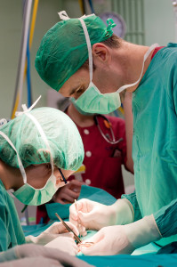
Lung biopsy should be performed by a cardiothoracic surgeon expert in VATS (video assisted thoracoscopic surgery). This means that two small holes are created and telescope-like equipment is then inserted into the chest. At least two and preferably three biopsies should be taken from three lobes of the lung on the same side. The tip of the middle lobe should not be sampled. Ideally, areas of advanced scarring are avoided. Areas of moderate disease are best.
Open surgery with a large incision is avoided to minimize pain and shorten recovery times. Most patients spend one night in the hospital. Surgery is done under general anesthesia. Immediately after surgery patients will wake up with a chest tube in their chest. This is generally removed the next morning.
What are the Risks of Surgery?
- Bleeding
- Infection
- Air leaks. This means that the lung is leaking air into the pleural space (area around the lung but inside the chest cavity). When this occurs, the chest tube must remain in place for longer.
- All the usual risks of general anesthesia (problems with putting a breathing tube into the airway such as injury to your teeth, allergic reactions to medicines)
Who Should Review Your Lung Biopsy?
Interpreting a lung biopsy is very difficult. Most general pathologists do not have the expertise to confidently tease apart IPF from fibrotic NSIP. As the treatments at present are so different for these two lung disorders, it is essential to get the interpretation correct. There are lung-specialized pathologists that are able to do this. I always send my lung biopsies to a national expert in interstitial lung disease to get a second opinion. This prevents misdiagnosis and treatment errors.